関連リンク
- Home
- イメージギャラリー
Select an application or technique:
最新イメージ
高速スキャンAFM
AFM / OT、先進光学
ライフサイエンス
ポリマー
ナノサイエンス
電気、磁気、熱特性測定
セルメカニクス、接着
単分子フォーススペクトロスコピー
ナノマニピュレーションとリソグラフィー
ラマン、TERS、SNOM
原子間力顕微鏡自動フォーススペクトロスコピー光ピンセット細胞/組織のメカニクスと接着
原子間力顕微鏡
NanoWizard® NanoScience AFM
-
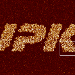
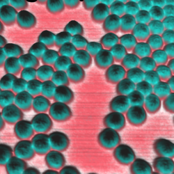
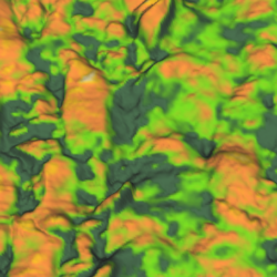
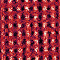
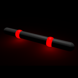
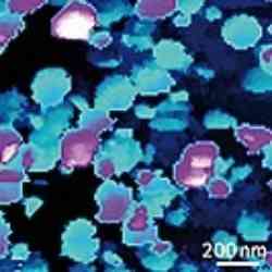
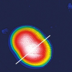
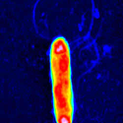
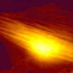
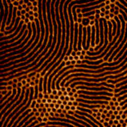
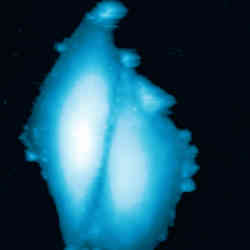
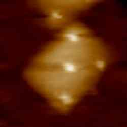
 MIPAM particle
 -
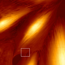
 JPK logo - piezo-response microscopy
 -
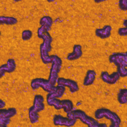
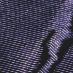
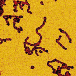
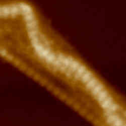
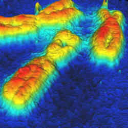
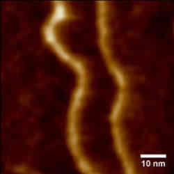
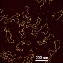
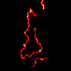
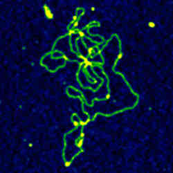
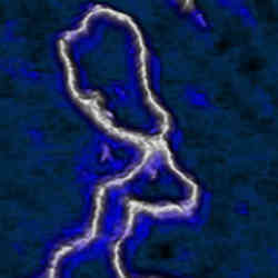
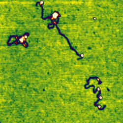
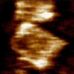
 DNA on octadecylamine
 -
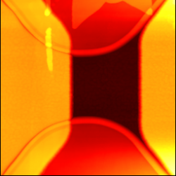
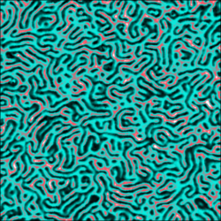
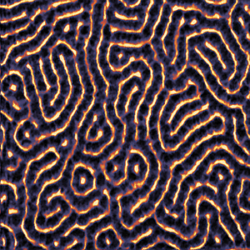
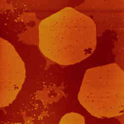
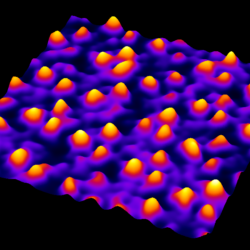
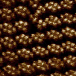
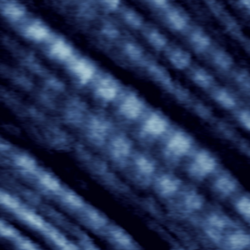
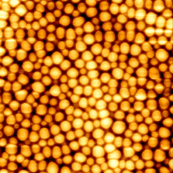
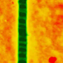
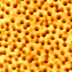
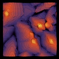 Pentacene layers
 -
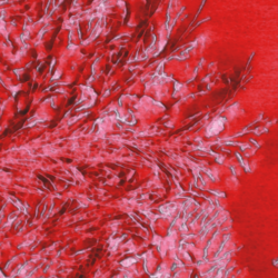
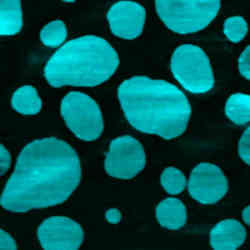
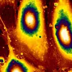
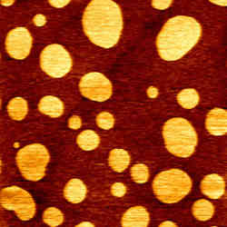
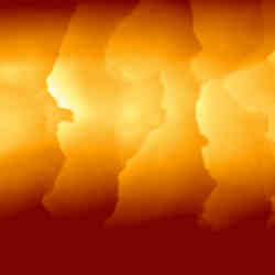
 Melting of a thin film of polystyrene
 -
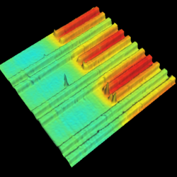
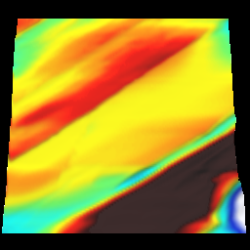
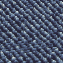
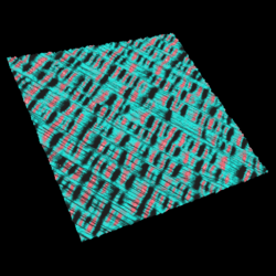
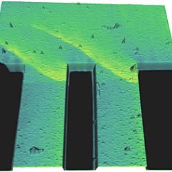
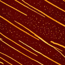
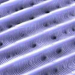
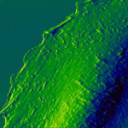
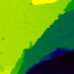
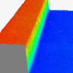
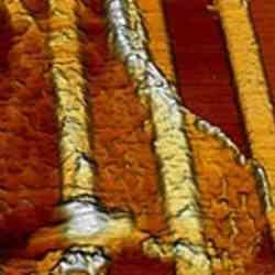
 Striped gold layer surface
 -
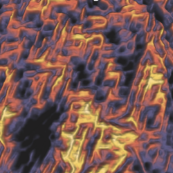
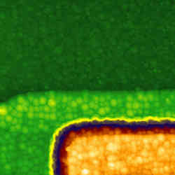
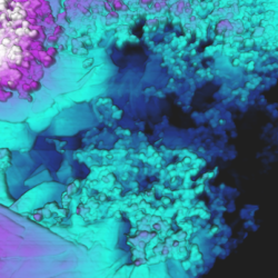 QI™-CAFM on battery electrode
 -
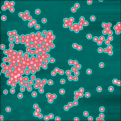
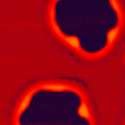
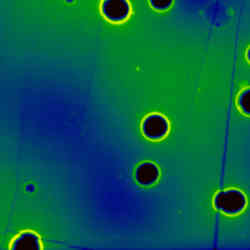
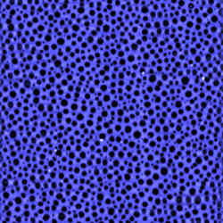
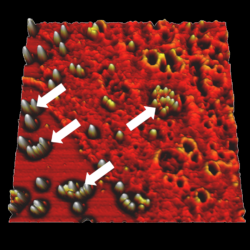
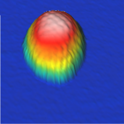
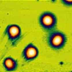
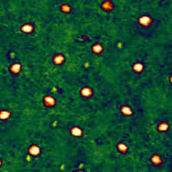
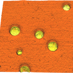
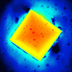
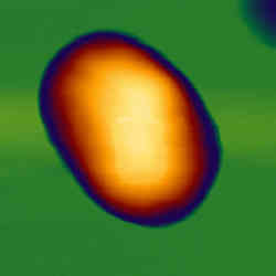
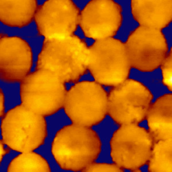
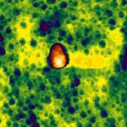
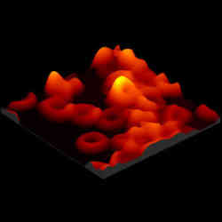
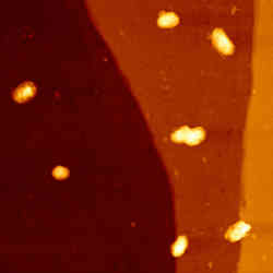
 QI™-EFM on NIPAM particles
 -
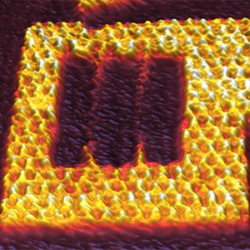
 QI™-KPM on an interdigitated electrode
 -
 Stretching of plastic Parafilm slide
 -
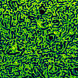
 Block-Co-Polymer thin section
 -
 Microrheology on silica beads
 -
 Stretching of plastic film
 -
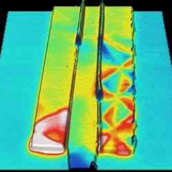
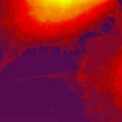
 Scanning thermal microscopy
 -
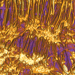
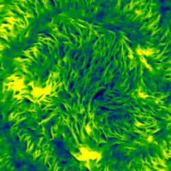
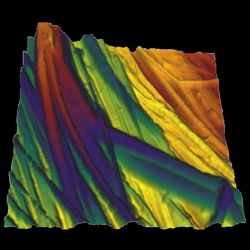
 PHB/V spherulite crystallization.
 -
 PFM lithograthy
 -
 CAFM on CU conduct layer
 -
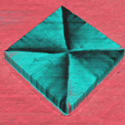 MFM on NiFe square structure
 -
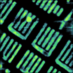
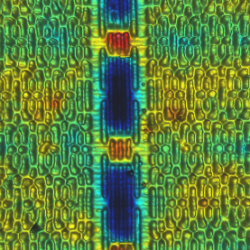 KPM on SRAM
 -
 Dendronized polymer
 -
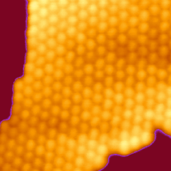
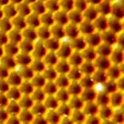
 Calcid - True atomic lattice resolution
 -
 Kraton
 -
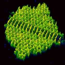
 Hexacontane
 -
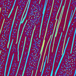
 Crystalline polyethylene at -120°C
 -
 Electrostatic Force Microscopy (EFM) on SRAM
 -
 Conductive QI™ measurement of a graphene flake
 -
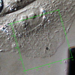
 Living KPG7 cell at 37°C - AFM and phase contrast
 -
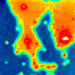
 Atomic resolution of mica taken on an optical microscope
 -
 Artificial polyprotein made of non-mechanical protein GB1
 -
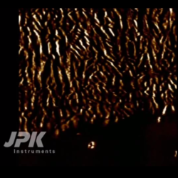
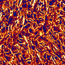
 PTFE (Teflon™) layer
 -
 MFM on hard drive
 -
 Kelvin Probe Microscopy (KPM) on an interdigitated electrode
 -
 Optically active conductive polymer film
 -
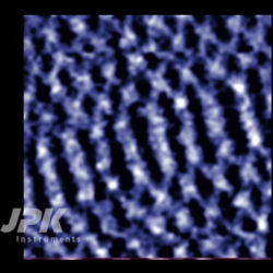
 STM of arachidic acid molecules
 -
 Electrical oxidation on silicon substrate
 -
 MFM images of rectangular magnetic structures
 -
 MFM images of a double T magnetic structure
 -
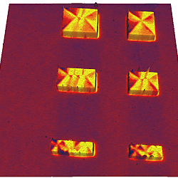 MFM images of six rectangular magnetic structures
 -
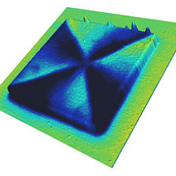 MFM images of a square magnetic structure
 -
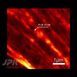
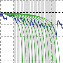
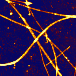
 Breaking point analysis on nano fibres
 -
 Hexacontane
 -
 Dendronized polymer - QI™ mode
 -
 Polystyrene-block-polybutadiene film with QI™
 -
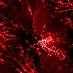
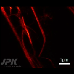
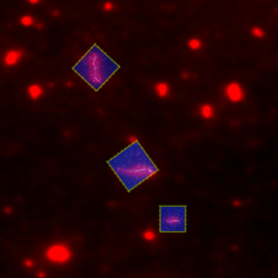
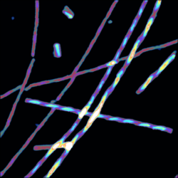
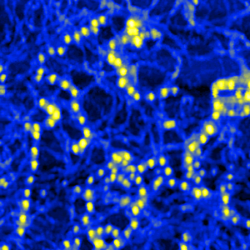
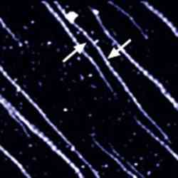
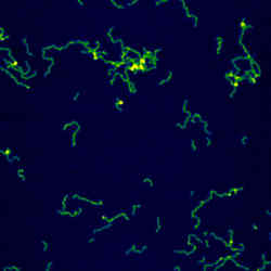
 Hexaphenyl nanofibers - AFM with fluorescence microscopy
 -
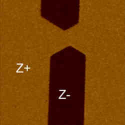 Domain visualization in lithium niobate crystal (PFM)
 -
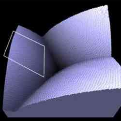
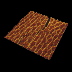
 Elastomeric polypropylene - 3D views
 -
 Hexaphenyl nanofibers
 -
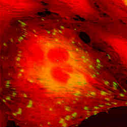
 Fluorescent polymer spheres - AFM with fluorescence microscopy
 -
 Polyelectrolyte shell - AFM with DIC
 -
 Hollow microcapsules
 -
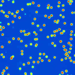 Polymer nanoparticles
 -
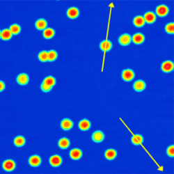 Polymer nanoparticle manipulation
 -
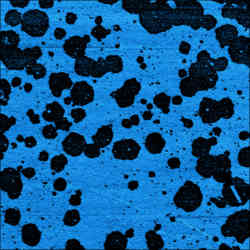
 Polystyrene surface with holes
 -
 PHB-PHV spherulites
 -
 SBS triblock copolymer
 -
 Langmuir blodgett film
 -
 Patterned PDMS polymer
 -
 Lithography on polymer
 -
 Polymer film
 -
 ITO on glass
NanoWizard® BioScience AFM
-
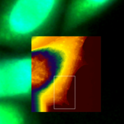
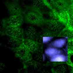
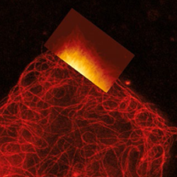 Living A549 cells - Correlative AFM and STED
 -
 Living Vero cells
 -
 DNA Origami at 150 lines/sec
 -
 VIDEO - Real-time rapture of microtubules - Correlative AFM and STED measurements
 -
 VIDEO - Real-time bending of microtubules - Correlative AFM and STED measurements
 -
 VIDEO - Stimulation of living fibroblast cells - Correlative AFM and STED measurements
 -
 Nanoruler - AFM and STED
 -
 Living A549 cells - Simultaneous AFM and STED
 -
 Astrocytes
 -
 Imaging of bacteria S-layer with QI™
 -
 Simultaneous AFM and STED of Living fibroblasts - Actin Filament Imaging
 -
 Simultaneous AFM and STED of Living fibroblasts - Microtubule Imaging
 -
 Cell/particle interaction - AFM with confocal microscopy
 -
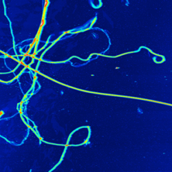
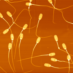 High-resolution imaging on sperm
 -
 Living CHO cell
 -
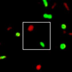
 Rad51 proteins bound to DNA - AFM with fluorescence microscopy
 -
 Fibronectin unfolding
 -
 DNA at -25 °C
 -
 CHO cell - AFM with phase contrast
 -
 QI™ DNA - Major and minor grooves
 -
 Recognition microscopy on biotin bead
 -
 Recognition on living keratinocyte cells
 -
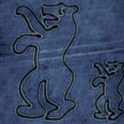
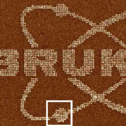
 DNA origami - faceman
 -
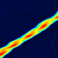
 Twisted insulin fibrils
 -
 Living CHO cells - AFM with fluorescence microscopy
 -
 Tendon tissue
 -
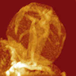
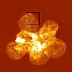
 Herpes Simplex Viruses
 -
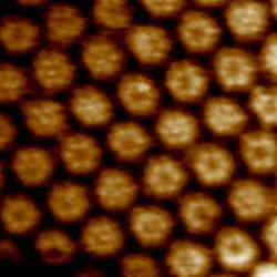
 Bacteriorhodopsin membrane - QI™ mode
 -
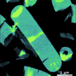
 Cell division E-coli bacteria
 -
 Human dental enamel
 -
 VIDEO - Live CHO cell dynamics
 -
 Tomato Bushy Stunt Virus
 -
 Plasmid DNA imaged in HyperDrive™ mode in buffer
 -
 KPG7 cell dynamics
 -
 Living Candida albicans - AFM with phase contrast
 -
 HeLa cell in buffer - AFM with STORM
 -
 Melting of lipid domains in buffer
 -
 Collagen in liquid, 70Hz line rate
 -
 Lambda phage DNA in liquid
 -
 Bacteriorhodopsin in buffer
 -
 Living fibroblast cell - AFM with phase contrast
 -
 Desulfobulbus bacterial cells
 -
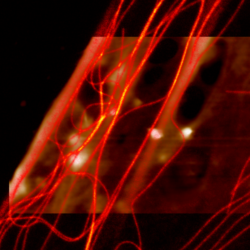
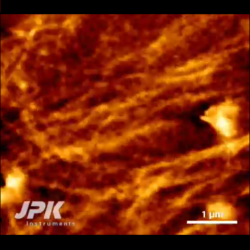
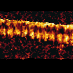
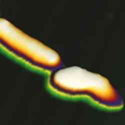
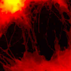
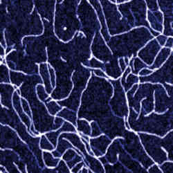
 Collagen fibres
 -
 Pea starch granules
 -
 Herpes Simplex Virus
 -
 Living Cyanobacterium
 -
 Living Escherichia coli bacteria
 -
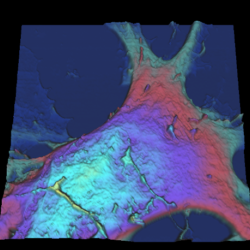
 Living dorsal root ganglion cells - AFM with DIC
 -
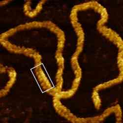
 Twisted amyloid fibrils
 -
 Collagen – AFM with phase contrast
 -
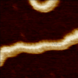
 Glucagon fibre
 -
 Fibrillin microfibrils
 -
 Malaria infected red blood cells
 -
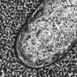
 L929 cell filipodia
 -
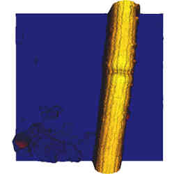
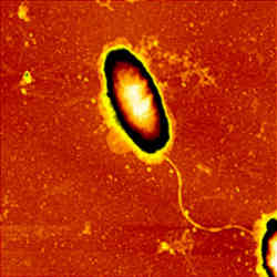
 Single waste water bacterium
 -
 OmpF protein crystal
 -
 Lipid bilayer - AFM with confocal microscopy
 -
 MDCK cells - AFM with confocal microscopy
 -
 Moth's eye
 -
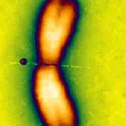
 Human lymphocyte chromosomes
 -
 Unfixed collagen in buffer
 -
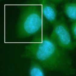
 Ptk2 cells - AFM with fluorescence microscopy
 -
 Living fibroblast cells – AFM with phase contrast and fluorescence
 -
 Living fibroblast cell
 -
 Lipid vesicles
 -
 Moth wing scale
 -
 Collagen manipulation
 -
 Gold clusters in water
 -
 REF52 cells – AFM with fluorescence
 -
 High-resoution image of HPI layer
NanoWizard® NanoOptics AFM
-
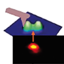
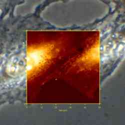
 Simultaneous AFM and FLIM measurements
 -
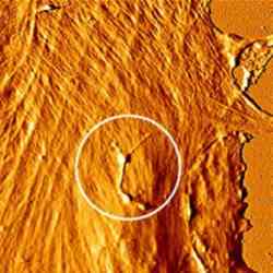
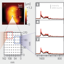 TERS mapping
 -
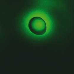
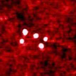
 Enhancement of fluorescence with gold nanoparticles
NanoWizard® ULTRA Speed AFM
-
 Correlative STED and AFM images of nanorulers
 -
 Correlative STED and AFM images of isolated sacculi of Bacillus subtilis
 -
 VIDEO - Living KPG7 fibroblast
 -
 VIDEO - High-speed imaging of DNA at 10 frames/sec
 -
 VIDEO - Melting and crystallization of a PLC thin film
 -
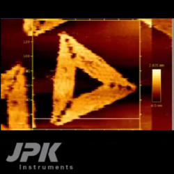 VIDEO - Triangular DNA origami
 -
 VIDEO - Soft DNA Origami in buffer
 -
 DNA Origami
 -
 VIDEO - PHB/V spherulite crystallization
 -
 Living cell dynamics
 -
 Celgard® fast scanning
 -
 True atomic resolution of mica
NanoWizard® Sense AFM
-
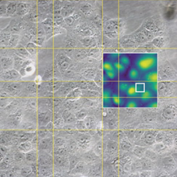
 Kelvin Probe Microscopy (KPM) on reference sample
 -
 Force mapping on elastomer
 -
 Contact Resonance frequency image of glas
 -
 DNA imaged with PeakForce Tapping mode
 -
 Piezoresponse Force Microscopy of a ferroelectric copolymer
 -
 Conductive AFM of a graphite-clay blend
 -
 E. Coli bacteria in buffer
 -
 Zeolite crystal
 -
 Diblock copolymer
 -
 Dendronized polymer molecule
 -
 Dendrimer molecules
 -
 HeLa cells in buffer
 -
 Living CHO cell
 -
 Xanthan molecules on mica
 -
 SAOS cells - AFM with confocal microscopy
 -
 Anodic aluminum
 -
 Structured PMMA surface
 -
 Fe-Pt nanoparticles in lipid bilayer
 -
 PPV viruses
 -
 DNA molecule on mica
 -
 Polystyrene surface with a failure crack
 -
 Phase-separated lipid bilayer
 -
 Quantum dots manipulation
 -
 SAOS chondrocyte cells – AFM with phase contrast
 -
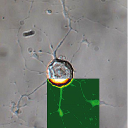
 Osteoblast cells with neuron
 -
 Polymer beads
 -
 Silicon calibration grid
 -
 PHB-PHV copolymer
 -
 Lambda-phage DNA in buffer
 -
 H. pylori bacterium
 -
 Erythrocyte with influenza viruses
 -
 Ceramic material for cell growth
 -
 Manipulation of a chromosome
 -
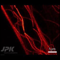
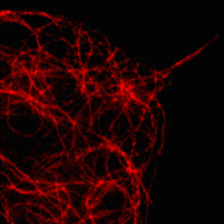
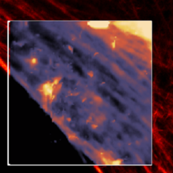
 Microtubules – AFM with fluorescence
 -
 Living neuron cell on chip
 -
 Erythrocytes
 -
 Human hair
 -
 DNA-nucleosome complexes
BioMAT™ Workstation
-
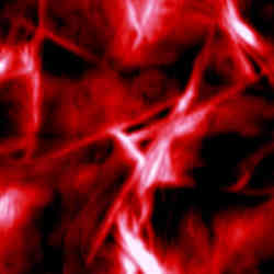
 J-aggregates
 -
 Paracoccus Seriniphilus bacteria
 -
 Living CHO on gold electrode
 -
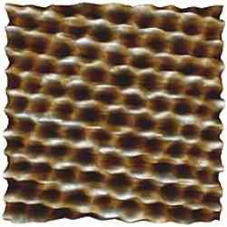
 Cow tooth – etched - AFM with upright microscopy
 -
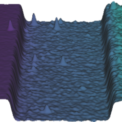
 Bacteria on pyrite surface - AFM with upright fluorescence microscopy
 -
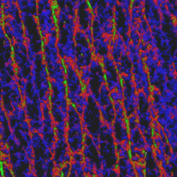
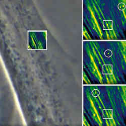
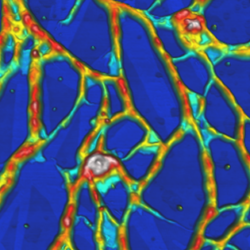
 Mouse cerebellum tissue - AFM with upright fluorescence microscopy
NanoRacer®
-
 VIDEO - Streptavidin binding to biotinylated DNA origami at 50 frames and 5000 lines/sec
 -
 VIDEO - High-speed imaging of DNA origami at 35 frames/sec
 -
 VIDEO - High-speed imaging of DNA at 50 frames/sec